Diagram Of Shoulder Ligaments - Anatomy Of The Shoulder Part 2 Ligaments And Capsules Mujo - Ebraheim's animated educational video describing the glenohumeral ligaments of the shoulder.the superior, middle, and inferior glenohumeral ligaments.
byAdmin•
0
Diagram Of Shoulder Ligaments - Anatomy Of The Shoulder Part 2 Ligaments And Capsules Mujo - Ebraheim's animated educational video describing the glenohumeral ligaments of the shoulder.the superior, middle, and inferior glenohumeral ligaments.. Both tendons and ligaments are dense regular connective tissue, because of its two properties: The goals of shoulder surgery are to reduce pain, increase function, mobility and stability of the joint, and correct deformities or injuries. Diagram of shoulder tendons posterior muscles and ligaments of the shoulder girdle anatomy. Shoulder ligaments labeled (page 1). Atlas of the anatomy of the joint of the shoulder on a ct arthrogram in axial, coronal, and sagittal sections, on a 3d images and on conventional athrogram.
Shoulder joint of human body anatomy infographic diagram with all parts including bones ligaments muscles bursa cavity capsule cartilage membrane for medical science education and health care. Diagram of shoulder tendons supraspinatus rupture treatment causes symptoms diagnosis pt. 17 photos of the diagram of shoulder muscles and tendons. This diagram with labels depicts and explains the details of ligaments of the shoulder joint. Click now and learn everything about its anatomy and function at kenhub!
Shoulder And Axilla Amboss from media-us.amboss.com Diagram of shoulder tendons posterior muscles and ligaments of the shoulder girdle anatomy. Start studying shoulder ligaments and tendons. 17 photos of the diagram of shoulder muscles and tendons. In the shoulder joint, the ligaments play a key role in stabilising the bony structures. Simple easy notes for quick revision for exams. Atlas of the anatomy of the joint of the shoulder on a ct arthrogram in axial, coronal, and sagittal sections, on a 3d images and on conventional athrogram. Ligaments and joints of female pelvis. (1) the collagen fibers are closely packed (dense) and leave relatively little open.
One or more ligaments provide stability to a joint during rest and movement.
Click now and learn everything about its anatomy and function at kenhub! Diagram of the shoulder ligament test setup. We've got the acromion posteriorly and the and lastly we've got this ligament called the coracohumeral ligament because it attaches from the coracoid process to the humerus. 6 describe briefly the abduction at shoulder joint. I've just switched over to this diagram here and we're looking at the same view, a lateral view of the right shoulder. These ligaments are main source of stability for the shoulder. This is an online quiz called shoulder ligaments. (1) the collagen fibers are closely packed (dense) and leave relatively little open. Ligaments and joints of female pelvis. Shoulder ligaments labeled (page 1). Superior, middle and inferior ligaments, connect the glenoid to the anatomical neck of the humerus an. Stretching or tearing them can make your joints unstable. The fixture included an additional aluminum plate (c) which was connected and moved with the instron actuator.
These tiny ligaments (with the acomioclavicular joint) play an important role in keeping the scapula attached to the clavicle and thus keeping your shoulder 'square'. However, one can recover the strength, and guard the shoulder ligaments by performing certain exercises. The shoulder is not a single joint, but a complex arrangement of bones, ligaments, muscles, and tendons that is better called the shoulder girdle. 17 photos of the diagram of shoulder muscles and tendons. The tighter the ligaments are, the less motion available.
Shoulder Joint Human Anatomy Arm Face Hand People Png Pngwing from w7.pngwing.com In anatomy, a ligament is a band or sheet of strong fibrous connective tissue that connects bones to other bones, or to cartilage, or supports an organ, such as the spleen, uterus, or eyeball. Shoulder ligaments labeled (page 1). The shoulder is not a single joint, but a complex arrangement of bones, ligaments, muscles, and tendons that is better called the shoulder girdle. Click now and learn everything about its anatomy and function at kenhub! The shoulder joint is supplied with blood by branches of the anterior and posterior circumflex humeral arteries diagram of the human shoulder joint, back view. Thompson shafts (a) and aluminum plates at the ends (b). Because the shoulder is a highly mobile joint, the ligaments must be loose to allow motion. (1) the collagen fibers are closely packed (dense) and leave relatively little open.
This is an online quiz called shoulder ligaments.
Atlas of the anatomy of the joint of the shoulder on a ct arthrogram in axial, coronal, and sagittal sections, on a 3d images and on conventional athrogram. Shoulder ligaments labeled (page 1). Because the shoulder is a highly mobile joint, the ligaments must be loose to allow motion. The shoulder joint (glenohumeral joint) is a ball and socket joint between the scapula and the humerus. Related online courses on physioplus. Ebraheim's animated educational video describing the glenohumeral ligaments of the shoulder.the superior, middle, and inferior glenohumeral ligaments. The shoulder is not a single joint, but a complex arrangement of bones, ligaments, muscles, and tendons that is better called the shoulder girdle. This is an online quiz called shoulder ligaments. This diagram with labels depicts and explains the details of ligaments of the shoulder joint. The left shoulder and acromioclavicular joints, and the proper. Shoulder joint is the most mobile joint of the human body. Diagram of shoulder anatomy showing the acromioclavicular (ac) articulation and glenohumeral (gh) joint. Thompson shafts (a) and aluminum plates at the ends (b).
These are the coracohumeral, glenohumeral and transverse humeral ligaments. This is an online quiz called shoulder ligaments. Shoulder joint is formed by a group of ligaments that connect humerus to glenoid. Stretching or tearing them can make your joints unstable. I've just switched over to this diagram here and we're looking at the same view, a lateral view of the right shoulder.
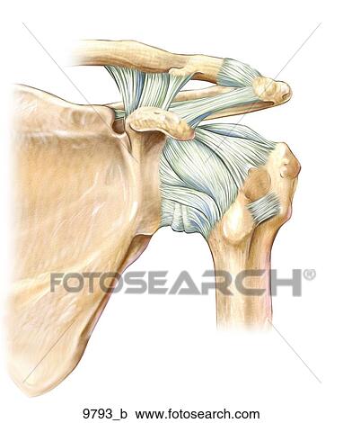
Left Shoulder Ligaments Anterior Unlabeled Clipart 9793 B Fotosearch from fscomps.fotosearch.com Thompson shafts (a) and aluminum plates at the ends (b). The fixture included an additional aluminum plate (c) which was connected and moved with the instron actuator. These are the coracohumeral, glenohumeral and transverse humeral ligaments. There are many shoulder ligaments which each play an important role in shoulder joint stabilization to various degrees: There is a printable worksheet available for download here so you can take the quiz with pen and paper. This is an online quiz called shoulder ligaments. Shoulder separation describes the condition in which the ligaments connecting the ac joint are injured and the acromion begins to move away from the clavicle. Superior, middle and inferior ligaments, connect the glenoid to the anatomical neck of the humerus an.
17 photos of the diagram of shoulder muscles and tendons.
We've got the acromion posteriorly and the and lastly we've got this ligament called the coracohumeral ligament because it attaches from the coracoid process to the humerus. Related online courses on physioplus. Things tend to wear out and break at the moving parts. The tighter the ligaments are, the less motion available. Lumbrical tendon passes volar to transverse metacarpal ligament. Simple easy notes for quick revision for exams. Because the shoulder is a highly mobile joint, the ligaments must be loose to allow motion. Atlas of the anatomy of the joint of the shoulder on a ct arthrogram in axial, coronal, and sagittal sections, on a 3d images and on conventional athrogram. 17 photos of the diagram of shoulder muscles and tendons. Shoulder separation describes the condition in which the ligaments connecting the ac joint are injured and the acromion begins to move away from the clavicle. The fixture included an additional aluminum plate (c) which was connected and moved with the instron actuator. Learn vocabulary, terms and more with flashcards, games and other study tools. Click now and learn everything about its anatomy and function at kenhub!
The coracohumeral, glenohumeral ligaments and the tendons of the supraspinatus and subscapularis muscles all serve to support and strengthen the joint diagram of shoulder. In the shoulder joint, the ligaments play a key role in stabilising the bony structures.